Multimodal Qdot (MQD) based nanoprobe for real time noninvasive bioimaging
PIs: Brij M. Moudgil, Glenn A. Walter, Edward W. Scott and Swadeshmukul Santra
PERC Team members: B. M. Moudgil, P. Sharma, S. C. Brown, A. K. Saha, A. Singh, G. Pyrgiotakis
Grant Number: NSF NIRT 0506560
In a number of tissues stem cells are known to be recruited from their respective niche's to repopulate or fuse with existent cells. However, the exact cellular signal and conditions that favor cell targeting and fusion are currently not well understood and is an area of intensive research. Tracking cell movement in real time in biological processes requires use of highly sensitive and photostable probes. Conventional organic fluorescent dyes and green fluorescent protein based labels are not suitable for long time monitoring since they lack photo-stability, which limits the time for which they can be tracked in biological processes.
There is substantial research and development interest in using fluorescent nanoparticles, e.g. dye-doped nanoparticles, fluorescent polymer microspheres, gold nanoparticles, quantum dots (QD), for bioimaging applications such as cell labeling, pathogen detection, early diagnosis of tumors etc. QD stand out amongst nanoparticle based labels because of their small size ( <10nm i.e. about 1000 times smaller than a typical cell); high photostability, making them suitable for real time, highly sensitive biological experiments, and are suitable for multiplexing (multiple labeling while using single excitation wavelength).
Optical imaging however provides limited anatomical background information, and also suffers from artifacts due to tissue absorbance and scattering. In this regard probes combining detection capability by optical and magnetic resonance imaging (MRI) are advantageous as they integrate the advantages of high sensitivity (from optical method of detection, e.g., fluorescence) with the potential of true three dimensional imaging of biological structures and processes at cellular resolution (MRI).
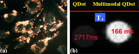 (a) Fluorescence emission from the MQD uptaken by the muscle derived stem cells and (b) Positive T1 contrast from MQD (on right) as compared to QD (without Gd(III)) as control (left) at 4.7T.
The objective of the project is to develop a multimodal QD-based nanoprobe that will be optically active to track cell delivery and will be further acted upon when a labeled stem cell fuses with tissue expressing β-galactose to generate MR contrast.
References
- P. Sharma, S. Brown, G Walter, S Santra and B Moudgil, " Nanoparticles for Bioimaging" Advances in Colloid and Interface Science, 123-126, 471-485, 2006.
- S. Santra, H. Yang, J. Stanley, P. Holloway, B. Moudgil, G. Walter, R. Mericle, "Rapid and effective labeling of brain tissue using TAT-conjugated CdS:Mn/ZnS quantum dots". Chemical Communications (Cambridge, United Kingdom) 25, 3144-3146, 2005
- H. Yang, S. Santra, G. Walter, and P. Holloway, "Gd(III)-Functionalized Fluorescent Quantum Dots as Multimodal Imaging Probes", Advanced Materials, 18, 2890-2894, 2006
- S. C. Grant, T. Zheng, G. P. Marshall II, H. Yang, P. Sharma, H. Cornnell, S. Santra, P. A. Holloway, B. M. Moudgil, E. W. Scott, E. D. Laywell, G. A. Walter, A. S. Edison, D. A. Steindler, M. D. Weiss, 'MR Microscopy of Multipotent Astrocytic Stem Cells Labeled with Multimodal Qdots Applied to a Neonatal Murine Model of Hypoxic Ischemic Encephalopathy' International Society for Magnetic Resonance in Medicine Meeting, May 2006, Seattle, WA. p. 1880.
- A. Y. Louie, M.M. Huber, E.T. Ahrens, U. Rothbacher, R. Moats, R.E. Jacobs, S.E. Fraser, T.J. Meade, 'In vivo visualization of gene expression using magnetic resonance imaging" Nature Biotechnology, 18, 321-325, 2000